MR-EYE: ultra high field magnetic resonance imaging of the eye

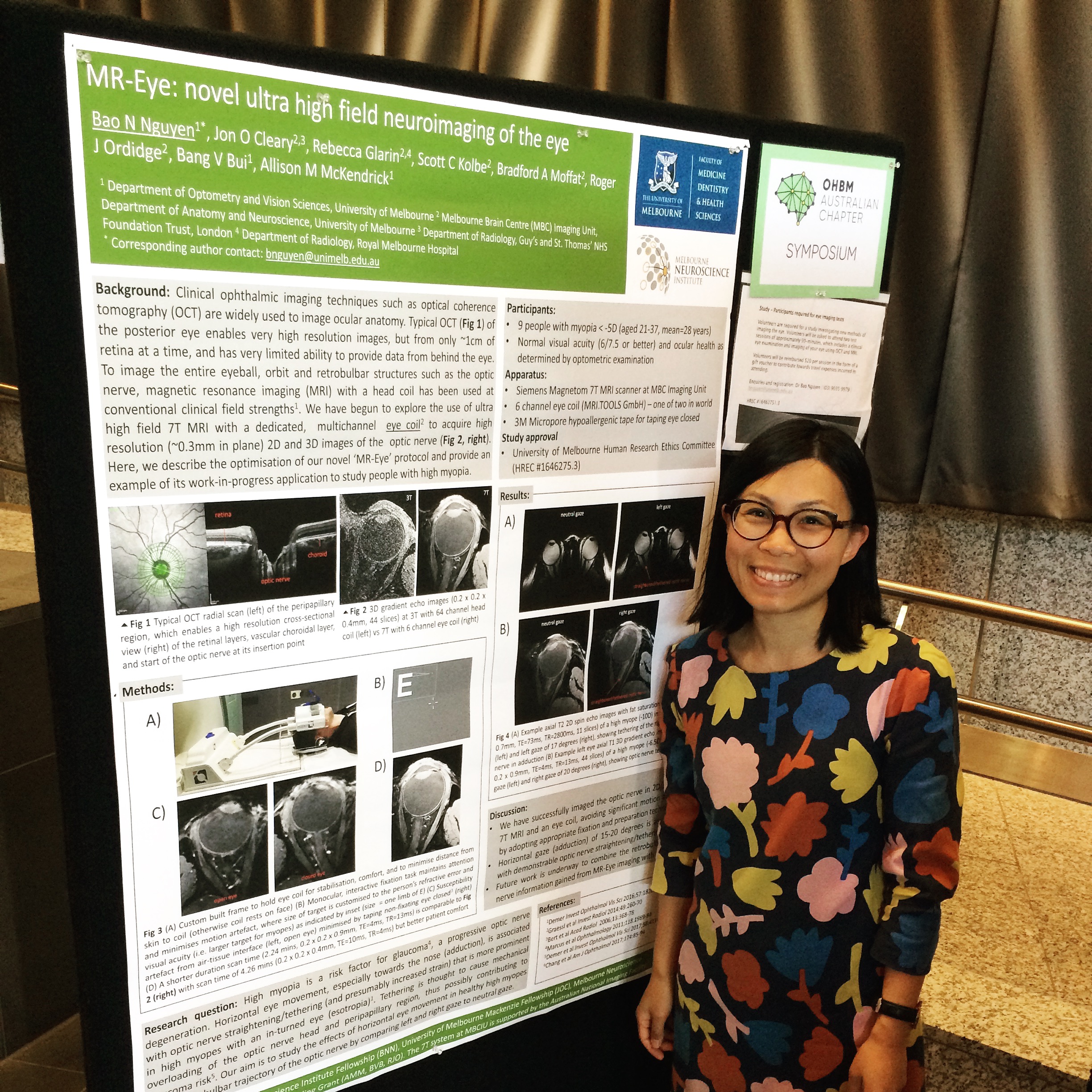
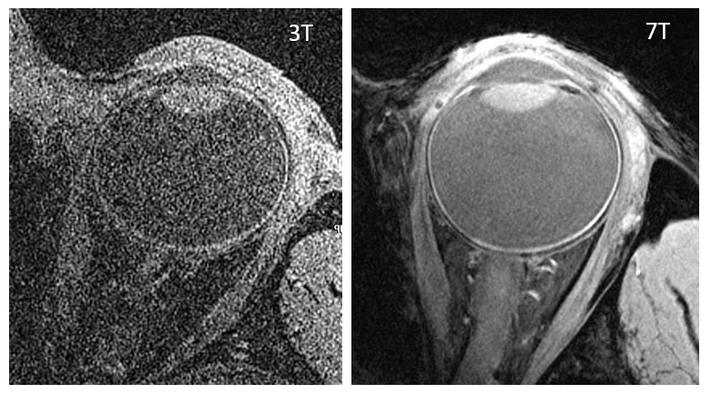
Thanks to a Melbourne Neuroscience Institute fellowship, Dr Bao Nguyen was able to spend a year pursuing a new collaboration between our lab and the Melbourne Brain Centre Imaging Unit at the University of Melbourne. The Imaging Unit houses a unique combination of a state-of-the-art 7 Tesla magnetic resonance imaging scanner (known as 7T MRI, which has improved signal-to-noise ratio compared with conventional clinical scanners of 1.5T and 3T) and a dedicated eye coil (not the usual head coil that is used for brain imaging) that is designed specifically for imaging the eye and surrounding anatomy.

Our collaboration has led to two papers. Firstly, we were invited to feature our work in a special issue of Magnetic Resonance Imaging Clinics of North America describing how we used the technology to acquire 2D and 3D images of ocular anatomy. We demonstrated that, with careful consideration of participant preparation and sequence selection to maximise image quality, it is clinically feasible to obtain ultra high field ocular and orbital MRI images with minimal artefacts. This information is useful for improved MRI investigations of ocular conditions that affect the structural integrity of the eye and its surrounding anatomy. The paper can be accessed here.

Example 3D MRI images (0.2 x 0.2 x 0.4 mm size, 44 slices) at 3T with a 64 channel head coil (left) versus at 7T with a 6 channel eye coil (right)

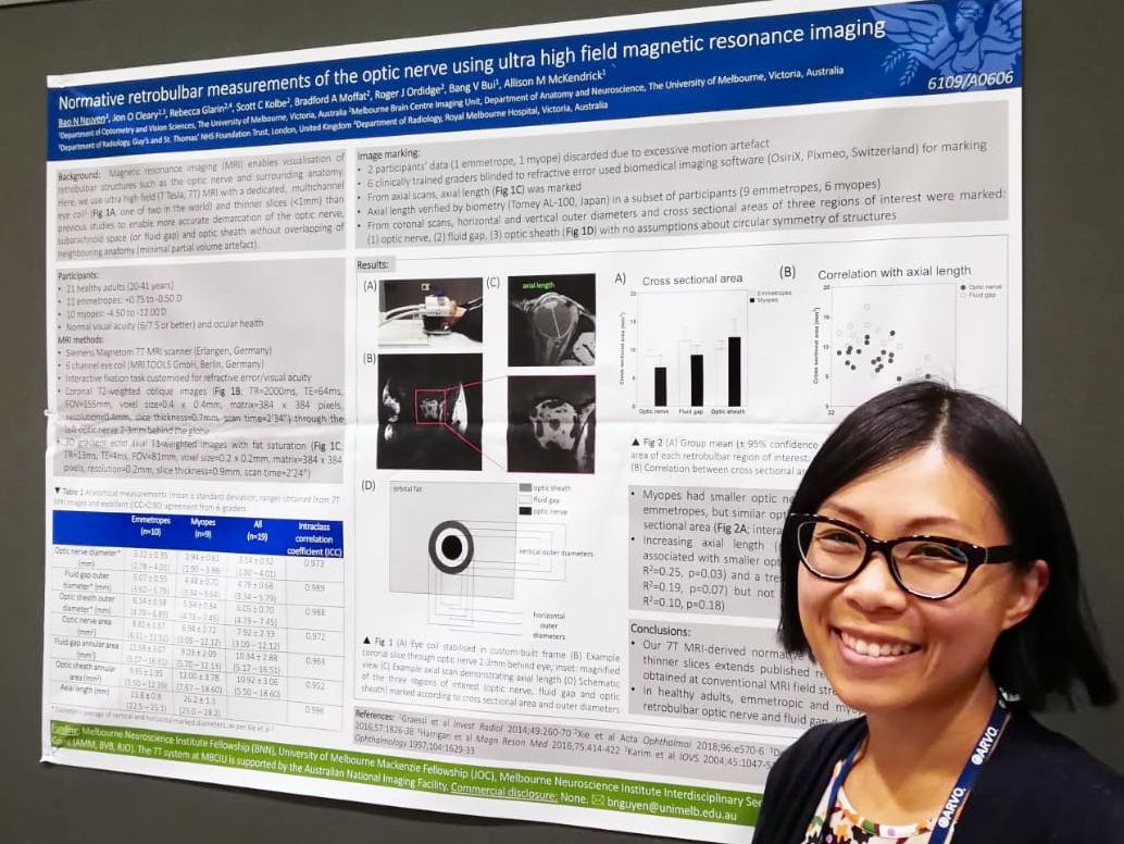
Our second paper, published in Translational Vision Science and Technology, demonstrates the feasibility of using 7T MR-EYE (with a dedicated eye coil) to enable measurement of the optic nerve, subarachnoid space (where fluid surrounding the brain also flows) and optic nerve sheath (the protective covering around optic nerve) behind the eye. The area just behind the eyeball, where the optic nerve exits the eye, is of clinical interest because this is where changes in intracranial (brain) pressure can impact on the optic nerve. It is also of clinical interest to better understand why some optic nerves may be more susceptible to damage, such as in eyes that are elongated (high short-sightedness, or myopia). Because one of the major advantages of MRI over conventional ophthalmic imaging such as optical coherence tomography (OCT) is the ability to image anatomy at depths behind the eyeball, we used our MR-EYE technology to image young, healthy eyes with and without high myopia to see if there were any anatomical differences. Our findings suggest that even without any signs of optic nerve damage, the cross-sectional dimensions of the optic nerve and fluid-filled subarachnoid space behind the eyeball are influenced by the degree of myopia. The paper is available to read open-access here.

7T MR-EYE can be used to take a ‘slice’ (see green line) through the optic nerve about 3mm behind the eyeball. The cross-sectional slice obtained can then be imaged to measure the cross-sectional area of the optic nerve, surrounding fluid-filled subarachnoid space, and surrounding optic nerve sheath

Part of this work has also been presented at several national and international conferences, including at the 2019 Association for Research in Vision and Ophthalmology in Vancouver, Canada (in the pre-COVID era!)

Dr Bao Nguyen presenting her MR-EYE poster at the 2019 Association for Research in Vision and Ophthalmology in Vancouver, Canada

MR-Eye: novel ultra high field neuroimaging of the eye

In October 2018, Bao presented a poster at the inaugural symposium of the Organisation for Human Brain Mapping Australian Chapter, which was held at the Melbourne Brain Centre at Parkville, Victoria.

The goal of the newly formed Australian Chapter is to bring together scientists and clinicians working in the fields of fundamental and clinical neuroscience and enable the maintenance of, and formation of, collaborations across all disciplines of neuroscience and neuroimaging.

An example of our interdisciplinary collaborative work, which Bao presented, is the development and optimisation of a novel magnetic resonance imaging (MRI) technique using a customised eye coil (only one of two in the world) that we have called ‘MR-Eye’. Most clinical MRI is conducted on a 1.5 Tesla or 3 Tesla machine. This imaging is special because it utilises the rare, stronger magnet (7 Tesla) imaging machine at the Melbourne Brain Centre Imaging Unit. The preliminary work has involved researchers from our laboratory, from our Department of Optometry and Vision Sciences, and from the wider community at the University of Melbourne and hospital institutions. We will be sure to keep you updated on the progress of this new neuroimaging technique!

04C5BA77-E426-43C2-867B-FA9DDDCA3253.jpeg

Bao Nguyen presented a poster at the inaugural symposium of the Organisation for Human Brain Mapping, titled: “MR-Eye: novel ultra high field neuroimaging of the eye”


Goodbye 2015… and welcome to the new year!

Happy new year! Our lab has returned in 2016 after a well deserved break while the University of Melbourne was closed for the festive season.

In 2015 we said goodbye to a few of our lab members:

  • Luke Chong and Jia Jia Lek graduated from their PhD degrees
  • Shonraj Ballae successfully completed his PhD thesis and is due to graduate from his degree
  • Juan Sepulveda graduated from his Masters degree
  • Siyuan Chen moved to France to continue her research

Janet Chan also successfully completed her PhD thesis and is due to graduate, and has returned to our lab as a post-doctoral research fellow.

We also have a new page as part of the University of Melbourne Department of Optometry and Vision Sciences website, where you can find out about our current research projects and collaborations

We have had some overseas visitors arrive in 2015 as part of our international collaborations. Dr Fumi Tanabe moved to Melbourne in October 2015 from the Department of Ophthalmology at Kinki University, Japan. She will be spending some time here continuing her research in glaucoma and visual field tests. At the end of 2015, Dr Quentin Lenoble from Universite Lille Nord, France, visited our lab for a few weeks as part of an international collaborative grant between our Australian and French research institutes.

Of course, we like to show our guests the Australian way of life and what better time of year than our summer: pizza picnics, walks on the beach, penguin spotting, indoor bubble soccer (this was our chosen end-of-year ‘lab event’) and one of our favourite outdoor sports, cricket.

fumi cricket

Fumi practises her batting skills with encouragement from Andrew, cricket enthusiast

phillip island

Fumi, Quentin, and Kabilan visit Phillip Island, home of the fairy penguins, on Christmas Eve

IMG_0479

Our lab end-of-year event where we attempted to play indoor soccer while in an inflatable bubble suit. There were lots of bumps, rolls, and upside-down moves but not so many goals!